How Muscles Work to Protect a Pitcher

Let’s talk about muscles. Muscles are the motors of the body. They are the components that generate movement. They can also absorb dangerous forces to protect more vulnerable tissues, like ligaments, and this is especially important for baseball pitchers.
Before I dive in, if you missed part one or two of the three part introduction to my views on the biomechanics of pitching, here’s a short summary.
I disagree with using the total elbow load as an approximation of the load on the ulnar collateral ligament (UCL). Therefore, I believe using the total load as an indicator of elbow injury risk is flawed.
One of the biggest drawbacks to using the total joint load is that it provides no information about the underlying muscles. This is why I account for the muscles, in addition to the ligaments and bones, when I analyze the biomechanics of a pitching motion using computational modeling techniques.
Now that you’re caught up, let’s focus on the muscles.
A muscle originates on one part of the body, at a location called its origin. It then crosses one or more joints and inserts on a different body part, at a location called its insertion. The body part of origin is typically the larger body part. When a muscle is excited by the nervous system, it contracts and attempts to shorten. In doing so, it exerts a pulling force at the origin and insertion. The pulling force causes the connected body parts to attempt to rotate about the encompassed joints in directions that shorten the muscle.
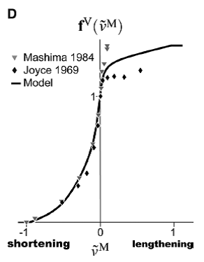
One of the most vital properties of muscle is the force-velocity property. Due to this property, the pulling force exerted by a muscle depends on the speed with which it is changing length. When a muscle is contracting and shortening quickly, it cannot generate much pulling force. This is called a concentric contraction. In contrast, when a muscle is lengthening but also excited by the nervous system (i.e. attempting to contract), it can generate a lot of pulling force. This is called an eccentric contraction. A stationary contraction, when the muscle is not changing length, is called an isometric contraction.
Figure from Millard et al. 2013 [1]. The vertical axis shows the multiplier by which the force exerted by a muscle is decreased or increased depending on contraction type.
An eccentric contraction occurs when an outside force is applied to a muscle that is greater than the maximum internal pulling force it can generate in its current condition. The maximum internal pulling force is governed by the muscle’s structure, its current level of excitation from the nervous system, and several other physiological factors. As seen in the figure, a muscle can actually produce more force during lengthening than it can when it is either shortening or stationary.
(A great reference for the previous paragraphs is this textbook from Dr. Rick Lieber [2].)
Our bodies often use eccentric contractions to decelerate body parts after periods of rapid acceleration, which is exactly what happens in the forearm during pitching.
Other examples of concentric, isometric, and eccentric contractions can be seen when a person is doing pull-ups. When the person is pulling up to the bar, his or her biceps muscles are concentrically contracting. When the person is holding at a steady position, the muscles are isometrically contracting. And finally, when the person is slowly lowering himself or herself down from the bar and controlling the descent, the biceps are eccentrically contracting.
Understanding muscle force production under all conditions is critical to analyzing and improving pitching mechanics, especially at the elbow. All of the muscles that cross this joint make essential contributions using all different types of muscle contractions.
And in baseball pitching circles, one group of elbow muscles gets more attention than the rest: the flexor-pronator muscles.
This muscle group gets more attention with good reason – studies have shown that muscle contractions in this group can relieve loading on the UCL [3-5]. Lesser loading on the UCL likely reduces the risk for a UCL tear and Tommy John surgery. Accordingly, injuries in this muscle group likely increase UCL loading during pitching and therefore may increase UCL injury risk.
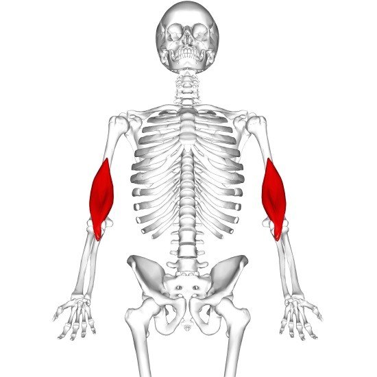
The flexor-pronator muscles are able to protect the UCL because they reside in similar locations to the UCL on the body [6]. Both the muscles and the ligament originate on the inside (the medial side) of the bone of the upper arm (the humerus). Thus, when the muscles are contracting, they are likely absorbing force that would otherwise be damaging the UCL.
The UCL originates on the humerus and then simply crosses the elbow joint to insert on the medial bone of the forearm (the ulna). However, the paths of the flexor-pronator muscles from origin to insertion are far more complex. In this muscle group, there are four muscles (FDS, FCR, FCU, and PT) that collectively cross the elbow, forearm, wrist, and even finger joints.
Try placing your left hand on the inside of your right elbow and then making a tight fist with your right hand. You will feel the movement of the tendons of your flexor-pronator muscle group with your left hand when you squeeze your right hand.
Because of the complexity of the flexor-pronator muscle paths, we cannot look at one arm joint or posture in isolation when we’re trying to analyze a pitcher’s elbow injury risk. Motions at the elbow, forearm, wrist, and even fingers all impact the outputs of the flexor-pronator muscles and their collective ability to protect the UCL. Depending on a pitcher’s mechanics and nervous system excitation patterns, the muscles can either be contracting concentrically, eccentrically, or even isometrically at any point in the pitching motion.
Therefore, an understanding of how a pitcher’s muscles are acting during his specific motion is imperative to assessing risk and prescribing effective training regimens.
This is exactly what I do in my biomechanical analysis approach [7]. I use a computational musculoskeletal model of the human body to compute and describe muscle actions, and then assess injury risk based on the results. From looking at the muscle actions, strategies can be devised that target the most important muscles in the most effective ways possible.
In the high school, college, and professional pitching motions I’ve analyzed, I’ve observed both concentric and eccentric contraction patterns in the flexor-pronator muscles during time periods of increased UCL injury risk. Different contraction patterns require different training techniques.
I do not agree with forcing a pitcher to conform to exact “ideal” mechanics. I do believe there should be mechanical guidelines, but I think it is essential to let a pitcher’s individual anatomy and muscle physiology guide his precise mechanical approach. Look at Greg Maddux. Look at Randy Johnson. Look at Pedro Martinez. Pitching success comes in all shapes and sizes.
Dr. James H. Buffi has a degree in mechanical engineering from the University of Notre Dame and a PhD in biomedical engineering from Northwestern University. His doctoral dissertation was called, “Using Biomechanical Modeling and Simulation to Calculate Potential Muscle Contributions to the Elbow Varus Moment during Baseball Pitching.” He has also been a visiting scholar in the National Center for Simulation in Rehabilitation Research at Stanford University as well as a visiting researcher at Massachusetts General Hospital. You can follow @jameshbuffi on twitter.
References:
- Millard, M., et al., Flexing Computational Muscle: Modeling and Simulation of Musculotendon Dynamics. Journal of Biomechanical Engineering-Transactions of the Asme, 2013. 135(2).
- Lieber, R.L., Skeletal muscle structure, function & plasticity : the physiological basis of rehabilitation. 3rd ed. 2009, Philadelphia: Lippincott Williams & Wilkins.
- Lin, F., et al., Muscle contribution to elbow joint valgus stability. Journal of Shoulder and Elbow Surgery, 2007. 16(6): p. 795-802.
- Seiber, K., et al., The role of the elbow musculature, forearm rotation, and elbow flexion in elbow stability: an in vitro study. Journal of Shoulder and Elbow Surgery, 2009. 18(2): p. 260-8.
- Udall, J.H., et al., Effects of flexor-pronator muscle loading on valgus stability of the elbow with an intact, stretched, and resected medial ulnar collateral ligament. Journal of Shoulder and Elbow Surgery, 2009. 18(5): p. 773-778.
- Davidson, P.A., et al., Functional-Anatomy of the Flexor Pronator Muscle Group in Relation to the Medial Collateral Ligament of the Elbow. American Journal of Sports Medicine, 1995. 23(2): p. 245-250.
- Buffi, J.H., et al., Computing Muscle, Ligament, and Osseous Contributions to the Elbow Varus Moment During Baseball Pitching. Ann Biomed Eng, 2014.
If you have not yet picked up a copy of ‘The Arm’ and you are the parent or coach of an athlete, picking up a copy is highly recommended.
Comment section
Add a Comment
You must be logged in to post a comment.


Why Overuse May Not Be Baseball's Problem for Pitchers - Driveline Baseball -
[…] for a pitcher to expose his or her muscles to controlled demanding situations where they can adapt. As I’ve said before, stronger muscles can generate and absorb more power. At joints like the elbow, stronger and more […]
Bullpens, Tracking Elbow Torque, and mStress - Driveline Baseball -
[…] Doesn’t account for muscles […]
Reviewing Offspeed Pitches and Elbow Torque - Driveline Baseball -
[…] also possible that those differences can change how the muscles in the forearm work. Leaving the overall torque similar but distributed in a different […]
Momentum and Arm Action: Myths, Misunderstandings -
[…] times before on this blog (this long article on Joel Zumaya’s elbow is a good start), the flexor-pronator mass dynamically stabilizes the elbow against valgus force and is thought to provide additional protection to the UCL during baseball […]